Mostrando las entradas con la etiqueta medical. Mostrar todas las entradas

domingo, 5 de enero de 2014

Dr. Couney and the Coney Island 'Child Hatchery'.


Theme Park History: Dr. Martin Couney and the Coney Island 'Child Hatchery'

Written by 
Published: October 20, 2013 at 10:25 AM

Of all the stories in theme park history (and perhaps medical history), one of the most curious has to be the story of Dr. Martin Couney.
Dr. Martin Couney
Born in 1870 in Germany, Dr Couney was one of the early pioneers of neonatology. He helped to develop the baby incubator and methods of caring for premature babies. In the late 1890s, his senior associates tasked him with spreading the word of the new technology to doctors and hospitals. Couney developed an exhibit and began demonstrations at fairs and expos around the world. The exhibits proved to be very popular, but more so with the curious general public than the medical industry they were intended to reach. The exhibit generated considerable crowds and revenue, but doctors and hospitals just weren’t that interested at the time.
After traveling exhibitions throughout Europe and the US for a few years, Dr. Couney set up a permanent exhibition in the newly opened Luna Park at Coney Island. In those days, hospitals had no special care for premature babies, so Couney was never short of patients. The outside of the building was no different than the other sideshows surrounding it. The sign above the door read “Life Begins With The Baby Incubator.” Customers were enticed in by a carnival barker and charged 25 cents to come and see the “child hatchery.”

Coney Island incubator exhibit
The inside was essentially a hospital. The atmosphere was quiet and clinical, incubators lined the walls, and trained nurses were employed to care for the babies. One of the nurses was Couney’s daughter, who ironically enough was born premature and spent some time in the incubator herself. The wet nurses employed to feed the babies were ordered on diets, and were fired if caught eating a hot dog or some other fried fare from the boardwalk. Tour guides were fired if they made jokes during the presentation. The rules and regulations for infant care were strictly enforced, and professionalism was emphasized. It was important to distinguish themselves at least a little from the pandemonium surrounding them.

Coney Island nurses Coney Island incubators
Naturally, there were those opposed to the idea of putting premature babies on display for the purposes of entertainment and profit. More than once there was a movement to shut him down. Dr. Couney had his reasons though, for throughout the show’s existence, he never charged a cent to the parents of the children he treated. It was the revenue of the paying customers covering the very high operating costs. He never took a payment for his services, and he accepted children of all kinds. Race, economic class, and social status were never factors in his decision to treat. The names were always kept anonymous, and in later years the doctor would stage reunions of his “graduates.” The medical profession that had once called him into question eventually embraced his methods and began promoting their use and sending him patients. Dr. Couney would eventually open more incubator attractions…a couple more at Coney Island and a handful around the country at other amusement centers and fairs.
Eventually, the enormous expense of running the exhibits began to outweigh the revenue as public interest in the attraction waned. Dr. Couney had made his case for the preemie though, and almost forty years after attraction opened, the first research center for premature infants was opened at Cornell University’s New York Hospital, reportedly differing very little from his operation. By this time, other hospitals were also opening treatment centers of their own. In 1943, Couney declared his work “finished” and closed for good. It’s reported that over 40 years of operation, Couney’s incubator attractions had an 80% success rate and saved about 6500 newborns from almost certain death. He died a few years later in 1950, having left his mark in both the theme park and medical industry.

lunes, 25 de marzo de 2013

An Ode to an Anatomical Venus: Waxing Poetic on the Uncanny Allure of 18th Century Dissectible Women


"Anatomical Venus," wax wodel with human hair and pearls in rosewood and Venetian glass case, "La Specola" (Museo di Storia Naturale), Florence, Italy; Probably modeled by Clemente Susini (around 1790)
An essay from collector, photographer, artist, and friend of the Atlas Obscura, Joanna Ebenstein of Morbid Anatomy about her love of the Wax Anatomical Venus.
Much of my artwork, scholarship, and work with the Morbid Anatomy Blog and Library revolves around the luxuriously bizarre Anatomical Venus, a kind of female wax anatomical model popularized in the 18th century. Over the past six years, I have made it my goal to find — and photograph! — as many of these amazing pieces as possible, and to learn as much as I can about these lovely ladies, with a special eye towards understanding the historical moment in which they rose to prominence as the ideal way to illuminate the anatomy of woman for a popular audience. I was recently invited to contribute an article on this topic to the "Enchantment" special issue of WSQ: Women's Studies Quarterly. Below is an excerpt from this piece, "Ode to an Anatomical Venus," and some of my photographs of these wax women. 
The "Venerina" or "Little Venus" anatomical model by Clemente Susini, 1782, as seen at the Palazzo Poggi in Bologna, Italy. Described on the museum website thusly: "The agony of a young woman is represented in her last instant of life as she abandons herself to death voluptuously and completely naked. The thorax and abdomen can be opened, allowing the various parts to be disassembled so as to simulate the act of anatomic dissection."
Clemente Susini’s Anatomical Venus, created around 1790, is the central object of my artistic and scholarly contemplation. She is, in my opinion, the perfect object; one whose luxuriously bizarre existence challenges belief. It — or, better she — was conceived of as a means to teach human anatomy without the need for constant dissection, which was messy, ethically fraught, and subject to quick decay. The Venus also tacitly communicated the relationship between the human body and a divinely created cosmos, between art and science, between nature and mankind as understood in its day.  
Detail of the ”Venerina" (Little Venus) anatomical model by Clemente Susini, 1782, Palazzo Poggi, Bologna, Italy
Referred to also as "The Demountable Venus," this life-sized, dissectible wax woman — who can still be viewed in her original Venetian glass and rosewood case at La Specola Museum of Zoology and Natural History in Florence, Italy, as well as in a number of other European museums — is adorned with glass eyes and human hair and can be dismembered into dozens of parts revealing, at the final remove, a beatific fetus curled in her womb. Her sisters — also anatomical models made under the artistic leadership of Susini, and referred to by such names as "The Slashed Beauty" and "The Dissected Graces" can be visited at a handful of European museums. Supine in their glass boxes, they beckon with a gentle smile or an ecstatic downcast gaze; one idly toys with a plait of real golden human hair; another clutches at the plush, moth-eaten velvet cushions of her case as her torso erupts in a spontaneous, bloodless auto-dissection; another is crowned with a golden tiara, while yet another has a silk ribbon tied in a bow tied around a dangling entrail. 

Anatomical model by Clemente Susini representing "deep lymphatic vessels in a female subject," human hair, wax, 1794, Museum of the History of the University, Pavia, Italy
Since their creation in late-18th century Florence, these wax women have seduced, intrigued, and instructed. Today, they also confound, troubling the edges of our neat categorical divides: life and death, science and art, body and soul, effigy and pedagogy, spectacle and education, kitsch and art. They are corporeal martyrs, anatomical odalisques, the uncanny incarnate. These wax models are the pinnacle of "artificial anatomies," a tradition of three-dimensional, anatomical teaching tools stretching back to the turn of the 18th century. The genre came into being around 1700 when Gaetano Giulio Zummo, known as Zumbo, accepted the commission of French surgeon Guillaume Desnoues to create a likeness of an important medical dissection that was beginning to decompose. Zumbo was a Sicilian abbot who delighted in the creation of wax miniature series “Theatres of Death” boasting names such as "The Plague" and "The Vanity of Human Greatness," and featuring exactingly rendered dead and tortured bodies. The product of Desnoues' and Zumbo's collaboration was the first wax anatomical teaching model, and established the tradition of an artistic/medical partnership in the creation of such tools.

"Slashed Beauty" Wax wodel with human hair and pearls in rosewood and Venetian glass case, "La Specola" (Museo di Storia Naturale), Florence, Italy;  Probably modeled by Clemente Susini (around 1790)
The Venus and her sisters were intended, from their very conception, not only to instruct, but also to delight and elicit the wonder of a popular audience and, beginning with their public debut in the 1790s, they did just that, attracting throngs of both local Tuscans and visitors on the Grand Tour circuit. Their popularity was so great that they ultimately inspired a series of knockoffs — first a series of similar models by the same workshop for Napoleon and Joseph II of Vienna and, later, in series of models, often advertised as "Florentine" or "Parisian" or even automated breathing Venuses that toured Europe, attracting masses of visitors to the popular anatomical displays found in Europe well into the 20th century. The uncanny allure of these somnambulant, neither-dead-nor-alive women was not lost on surrealist artists such as Paul Delvaux — who cited his visits to the Spitzner Collection (as seen in his painting "Le Musee Spitzner" of 1943), with its famous breathing Venus as a life and art-changing moment — and Marcel Duchamp, whose enigmatic peepshow Étant donnés seems to have been inspired, at least in part, by the paradoxes embodied by such figures. 

"The Slashed Beauty"Wax model with human hair in rosewood and Venetian Glass case; Probably modeled by Clemente Susini (around 1790), "La Specola" (Museo di Storia Naturale), Florence, Italy
At the time these models were conceived of and created, the anatomy of woman was of great interest, in both the medical and social arenas; the residue of these concerns, as argued by scholar Ludmilla Jordanova in Sexual Visions, can be found in the Anatomical Venus and other anatomical waxes of the era. The anatomy of woman was understood at this time to be the exception to the canonical body of the male: problematic, erratic, and troubling. It was also understood to be intimately tied to the female temperament, which was thought to be sensible (ie, sensitive), nervous, passionate, childlike, passive, and prone to such nervous disorders as hysteria — literally, "wandering womb." Men, in contrast, were understood to be muscular, vigorous, and reasonable. This difference is reflected in the models from the workshop of Susini. Each of his Venuses features a fetus — the raison d’être of woman, after all! — at the last stage of anatomical striptease. And, while male figures can be represented standing or reclining — and more often than not were portrayed completely skinned, demonstrating, say, human musculature — all the female figures are reclining and with their hyper-perfect skin intact, except for the places where the anatomical elements are exposed. The female figure, then, always remains beautiful and, one could argue, sexually desirable, and it is the line between her classic, serene beauty and the abjectness of her innards that adds to her special frisson.  

"Anatomical Venus" Wax wodel with human hair and pearls in rosewood and Venetian glass case, "La Specola" (Museo di Storia Naturale), Florence, Italy; Probably modeled by Clemente Susini (around 1790)
This frisson seemed to be a formula for popular success, so it is no surprise that Susini’s Venus and her sisters were not the first to employ it. Indeed, in 1719, Desnoues, the French surgeon who partnered with Zumbo in the creation of the first anatomical model, exhibited to the public a wax dissectible anatomized woman featuring a newborn child with the umbilical cord still attached. Fourteen years later, the Paris-trained anatomist, surgeon, and modeler Abraham Chovet exhibited in London ‘‘. . . the representation of a woman big with child chained upon a table; supposed to be opened alive. In the face there is a lively display of the agonies of a dying person, the whole body heaving and the hands clinched, the action suitable to the character of the subject.’’ This ingenious piece demonstrated the circulation of the blood during pregnancy via an network of blown glass tubes coursing with blood-red claret. 
You can read the entire "Ode to an Anatomical Venus" piece by clicking here
This piece could simply not exist without the wonderful work of scholars Roberta Ballestriero, Alessandro Riva, Lucia Dacome, Kathryn Hoffmann, Ludmilla Jordanova, Martin Kemp and Marina Wallace, Anna Maerker, Rebecca Messbarger, and Roberta Panzanelli. A much more detailed bibliography and list of citations can be found in the article itself. I am entirely indebted to their work in all of my research on this topic.
"Anatomical Venuses" Wax models with human hair in rosewood and Venetian glass cases; Workshop of Clemente Susini of Florence, 1781-1786 The Josephinum, Vienna, Austria

A Collection of
Curious Facts
© 2012 Atlas Obscura. All rights reserved.

jueves, 20 de septiembre de 2012

The Story of the Stone Child of Sens.


A lithopaedion, or stone-child, is a dead fetus, usually the result of a primary or secondary abdominal pregnancy, that  has been retained by the mother and subsequently calcified. This is the story of the earliest known case of this
phenomenon. It was discovered in 1582, at the autopsy of a 68-year-old woman in the French city of Sens, and described in a thesis by the physician Jean d'Ailleboust. The woman had carried her lithopaedion for 28 years. The ultimate fate of the lithopaedion specimen, which was widely traded throughout Europe in the 1600s before finally ending up in
Copenhagen, is traced.


INTRODUCTION
On May 16 1582, in the city of Sens, the 68-year-old Madame Colombe Chatri breathed her last. She was the wife of Loys Carita, a tailor, described as being of small stature, but otherwise 'bien forme & corpulent'. Twenty-eight years earlier, Madame Chatri had, for the first time, shown signs of pregnancy.

 Her menstruations ceased, her breasts swelled, her stomach increased in size, and she could even feel the child move within her. Some time before her birth was due,
Colombe Chatri was seized by violent labour pains. A great quantity of amniotic fluid, tinged with blood, was passed.
In spite of the predictions of the Sens midwives,


however, there was no childbirth; instead, her labour pains ceased, the movements of the child could no longer be felt, and her breasts diminished in size. Afterwards, Madame Chatri felt quite unwell, and she had to remain in bed for three full years. She could feel a hard tumour of considerable size, situated in the lower abdomen. Until the end of her life, she complained of tiredness, abdominal pains and loss of appetite. Only by means of provoking the appetite with herbs and vinegar sauces could she eat anything at all. When
gossiping with her neighbours in the street, they talked about her strange obstetrical mishap, and there was much speculation that she still had a fetus within her, and that it would kill her one day. Madame Chatri and her husband
consulted several physicians and surgeons, but none of them
could suggest a cure.


THE STONE-CHILD
In 1582, at the age of 68, Madame Chatri was described as being broken down by disease and old age. She died that year, and since there had been much gossip about her mysterious pregnancy many years ago, her husband requested that her body was to be dissected by two skilful surgeons, Claude le Noir and Iehan Couttas.
They cut through the stomach and peritoneum, and viewed the prodigious growth, which was wrinkled and formed like a turkey's crest. It was hard and brittle like a shell, and covered with what seemed like scales. The surgeons
'plunged their razors into it', but without being able to penetrate the hard shell. After wearing out the edge of their knives on the hard tumour, they fetched mauls and a drill, and finally succeeded to break it. They felt the head and right shoulder of the lithopaedion, but it was not until they had broken off a large portion of the covering shell, and seen the wonderful sight inside, that they understood what they were dealing with.
They ran to fetch some physicians, Jean d'Ailleboust among them. He could see a glimpse of the lithopaedion, which was covered by detritus and the remains of its inner membranes, and was as astonished as his surgical colleagues.
All the time, curious townsmen came running in to see this prodigy. The surgeons were busy telling the story, and to demonstrate the infant more clearly, they grasped the opening in the calcified shell with their iron hooks to tear it apart. After tearing with all their force, they broke it open,
and took out the lithopaedion, which they set out to dissect further. This was done with great haste, and Jean d'Ailleboust deplored that they had made it impossible to study closer the anatomy of the calcified shell and the nourishing vessels. The shape of the lithopaedion was roughly that of its rounded, calcified shell. The knees were bent, and the legs drawn up towards the chest. The feet and lower legs were fused by the calcific deposits. It could clearly be seen that the fetus was of the female sex. The head was lightly tilted to the right, and supported by the left arm.
The right arm extended down towards the navel: its hand had been broken off through carelessness when the lithopaedion was extracted. The bones of the head were transparent, and the fontanelles were not closed. The skin of the head was partially covered with hair. The lithopaedion had one sole tooth, situated in the lower jaw.


Not long after it had been delivered, the stone-child of Sens became one of the foremost curiosities of France. People traveled hundreds of miles to see and admire it. Jean d'Ailleboust needed no encouragement to write a thorough
Latin pamphlet about it, detailing the case history of Madame Chatri as well as the autopsy findings1. It was published in 1582, by the Sens printer Jean Sauvine, and soon became a medical best seller.


He also supplied a curious drawing (Figure 1) of the lithopaedion and its 'mother'. It is believed to depict the corpse of Columba Chatri lying on a richly padded bed, her abdomen dissected to show the lithopaedion in situ within its
calcified shell. Beside the bed, the lithopaedion is laid out on a pillow.  This practice was not unknown at the time:

 Although Jean d'Ailleboust was very unwilling to part from his great treasure, it is recorded that, in the late 1500s, it was purchased by a wealthy merchant, Monsieur Prestesiegle, who put it in his famous private museum of curiosities in Paris. It was examined there by Madame Louise Bourgeouis, the leading French midwife of her time5. Later, the lithopaedion was purchased by Etienne Carteron, a wealthy Paris goldsmith. He sold it, in 1628, to Gillebert
Bodey, a jewel merchant of Venice.  In the early 1650s, King Frederick III of Denmark was building up a large cabinet of curiosities at his castle in Copenhagen. In 1653, Frederick III bought the stone-child from its owner.


 The document of sale and certificate signed in 1628 were also turned over to the King, as well as a hand written copy of Jean d'Ailleboust's autopsy report, with a slightly different illustration (Figure 3): they are still kept in the
Royal Library of Copenhagen (Gl kgl Saml No. 1641). The sum paid was a well-kept secret, but it is unlikely that the Franco-Italian merchant let the King have it cheaply, particularly since he had himself paid a very high price for it.  A few years later, Thomas Bartholin described the lithopaedion closer in one of his collections of anatomical anecdotes9. By this time, it had become much the worse for wear, and it is likely that during its years in private hands, the lithopaedion had not always been treated with the reverence due to its age and fragility. Both arms were now broken off, the jaw was injured, and the skin
lacerated (Figure 2b). 


In the illustration (Figure 2c), the miserable-looking specimen is depicted sitting lopsidedly on a box. Part of the missing arm had been refastened, but otherwise, the lithopaedion was unchanged since it was described by Thomas Bartholin. In Jacobsen's 1710 catalogue10, it was more thoroughly described. The skin was now missing in large parts, and where it remained, it was quite black, giving the lithopaedion a strange aspect, as if it had been partly
dressed. 


In the 1820s, the Danish government decided to dissolve the Royal museum, and many preparations were scrapped or sold by public auction. Many bizarre pieces changed hands under these circumstances; among them the hand of a mermaid, solemnly described by Thomas Bartholin 170 years earlier, and an egg allegedly laid by a Norwegian peasant woman12. The lithopaedion was not among the preparations sold or thrown away, probably because it was still considered valuable. In 1826, it was transferred to the Danish Museum of Natural History, along with several other human, animal and vegetable specimens. In the late nineteenth century, the remaining exhibits from the Danish Museum of Natural History were transferred to the Zoological Museum of Copenhagen University, and a good many zoological specimens are still there. However, the lithopaedion had disappeared somewhere along the way,
together with the other medical curiosities from King Frederick's museum, among them a dicephalic child preserved in spirits, and a minute fetus, alleged to be one of the 365 children of the prolific Dutch countess Margaret
of Henneberg. It may well be that the lithopaedion of Sens was scrapped at this time, along with the other older medical specimens, of which no trace remains. An extensive search for them in the existing Danish museums has been fruitless.


A beautiful Latin poem was written by Jean d'Ailleboust in 1582 to celebrate the lithopaedion of Sens. He recalled the classical myth that after the Flood, the world was repopulated by the two survivors, Deucalion and Pyrrha, who walked the earth, throwing stones behind them, which, on striking the ground, became living people:


Pinxit Deucalion saxis post terga repulsis
Ex duro nostrum marmore molle genus:
Qui fit ut infantis, mutata sorte, tenellum
Nunc corpus saxis proxima membra gerat!


From the rocks Deucalion had dropped behind,
was fashioned the living flesh of humankind:
How was it then done, that a tender babe well formed


Information via:

"The earliest known case of a lithopaedion"
Jan Bondeson MD LicSc
J R Soc Med 1996;89:13-18

lunes, 17 de septiembre de 2012

Stone People: living statues.

In the 17th century the French physician Patin described the case of a local woman who had ‘turned to wood’. This ‘wood’ was actually bone and the woman possessed an incredibly rare condition that caused her muscles to be slowly turned to bone.

The earliest and most well documented case of fetal ossification dates back to 1582 when Mme Colombe Chatri died at the age of sixty-eight, and a twenty-eight year old fetus was removed from her womb. The "Stone-Child of Sens" should have been born in 1554; however, labor came and went with no delivery and in the resulting decades the fetus calcified and ossified within the womb, which actually formed a shell. Mme Chatri seemed to have lived a normal life, with the exception of regular abdominal pains. There will be more on the Stone Child in a subsequent post!


Fibrodysplasia Ossificans Progressiva or FOP, as the condition is know by today, affects 1 of 2 million people with varied severity. The condition is a genetic mutation in which the bodies of those affected cannot switch off the mechanism that grows the skeleton in the womb. Also, any small injury to connective tissue - muscles, ligaments, and tendons - results in the formation of hard bone around the damaged site. As it is a spontaneous genetic mutation, a FOB child can be born of normal parents – however persons with the condition have a 50% chance of passing the traits on to their offspring. As the hereotopic (extra) bone growth only becomes painfully obvious after a few years – 10 being the average - the only sign of the condition observable in an infant is malformed big toes. It is not until the second skeleton begins to form and mobility becomes severely restricted that the condition becomes evident.

Persons with FOP have been involved in sideshow and curio displays for centuries. Those with the condition were commonly called ‘ossified men’ or ‘stone men’, ‘The Ossified Man’ became a popular attraction. An ossified woman named Miss Emma Shaler once even shared billing with Harry Houdini in 1894. Strangely enough the ossified individual became quite a common attraction – likely due to the fact that it was a Marvel easily faked.

For those with the condition, life was far from easy. Movement was severely hampered and, in many cases, movement involved little more than lips and inner workings. The money these ossified men and women earned while on display paid for much needed medical attention. Many were often attended to during display by hired nurses. Few were able to eat anything, and their jaws became fused, and many had to sustain themselves on liquid diets. Thus many individual with the condition appeared incredibly gaunt and sickly. Most died quite young of pneumonia or other ailments that fed upon the sickly.

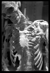
Harry Raymond Eastlack (pictured above) was born in the early 1930’s and was one of the last modern ossified men presented as a curiosity. He died of pneumonia in 1973, and his case is particularly notable because shortly before his death, he made it known that he wanted to donate his body to science. The gesture was in the hopes that in death he would be able to help find a cure for this rare and somewhat cruel disease. As per his wishes, his preserved skeleton now resides in The Mütter Museum in Philadelphia. Mr. Eastlack remains the best documented Stone Man in history.

Another FOP skeleton, belonging to a veteran Peter Cluckey, lives at the National Museum of Health and Medicine on the Walter Reed campus in Washington.
 

From public affairs officer Steven Solomon and my friends at the Kircher Society:


Peter Cluckey was born in 1882, enlisted in the Army at age 17 just after the Spanish-American War, retired from the service after 3 years, and rejoined in 1904. Two months after his second enlistment he experienced joint pain and stiffness after a horseback mounted drill held in cold rain. 

After several medical examinations he was diagnosed with “rheumatism chronic, articular, affecting both hips, knees, and ankle joints, and the right elbow.” Over the next 20 years his condition worsened to the point where every joint in his body became fused. Cluckey was moved into a sitting position so that he could be placed in a chair or on his side in bed to sleep. His front teeth were removed so that he could be fed soft foods. In his will, Cluckey donated his body to the museum and his skeleton has been on display seated in a wooden chair in the museum since his death in 1925.


We invite you to visit this fascinating specimen in person. Admission and parking are free.


Steven Solomon


Public Affairs Officer

National Museum of Health and Medicine

202-782-2200

www.nmhm.washingtondc.museum

jueves, 5 de julio de 2012

A Return to the Sepulchural Doctor's Shack

 An excerpt from Gakuranman's account of exploration of an old Medical shack in Japan:

A Return to the Sepulchral Doctor’s Shack

| Haikyo / Ruins |
 
Just shy of a year ago I came across an age-old medical shack hidden in the heart of the Japanese countryside. Brimming with mystery and intrigue, I thought I had seen everything the place had to hide, but a repeat visit with a fellow urbexer unearthed an abundance of creepy new discoveries. Just in time for Halloween…


The Doctor’s Shack, as I’ve nicknamed it, is a an old medical clinic abandoned around 60 years ago dating back to pre World War 2. The fact that a wooden building has survived this long along is quite remarkable in itself, but the shack is filled with a plethora of old medicine bottles and books labelled in German, likely harking back to the Meiji Period around 1870 when the government adopted German systems of medicine. According to Jikei University, one of the hallmarks of the German system was an “authoritarian discipline and an emphasis on research over treatment: patients were usually regarded as objects or even as raw material for medical research.”


My haikyo partner Florian pointed out a dusty, cold gynaecologist’s chair resting unceremoniously in the corner of the room that I had missed the first time around. Among the various containers filled with strange substances and toxic powders, were also rusty cutting instruments and old vessels for dispensing liquids. Literature scrawled in German text was strewn all over the upper floor of the building. The photo below of Thrombogen appears to be the German term for Thrombin, a drug administered to stop bleeding.


These were the sort of items I was consumed with photographing on my first visit. The sight of medicines that could be as much as 100 years old piked my curiosity and I was overtaken with an intense longing to know everything about its history, as well as being very concious of how fragile the place was. Perhaps this is why I overlooked much of the darker side of the Doctor’s Shack my first time around. The only thing I remember feeling an uncomfortable awareness of was the creepy deck chair tucked away at the end of a corridor on the second floor. But this was to be the first of many eerie, dark sights…


Florian was busy shooting the lower floors of the building, so I creaked up the staircase to the second floor to get some interior shots while the light was still good. The first time I had visited, the sun was already beginning to set and it had soon become pitch black inside causing me to miss the bizarre discovery below.


A footprint, or half of one had been left bleached into the woodwork. But that’s not the freaky part – this half-footprint was on the ceiling
I considered how it might have gotten there. Kids playing a trick? The shape of the foot was very pronounced and it was not made by any sort paint or ink; the mark was stained into the wood. Even if someone had played a tried and somehow managed to put a footprint on the ceiling, why was it only half a footprint?? Examining the photo above, the footprint cuts off where it meets the next wooden slat, leading me to think that perhaps the slat had been flipped upside down and that the other half of the print was on the floor above.
But there was no floor above…


Batting off numerous humming mosquitoes, I walked underneath the mark and continued down the hallway to take another look at the deck chair. I don’t quite know what it is about the thing, but staring at it sitting right at the end of a tiny corridor really creeped me out. Something, something sepulchral, held me back…
I could still hear Florian snapping away downstairs and cursing every now and then at the swarms of insects defying the colder weather and hunting for our blood. Another famous sight within the shack caught my eye in the Doctor’s bedroom.

It reads:
「健全ナル国民ノ育成ハ師表タル者ノ徳化ニ俟ツ。コトニ教育ニ従フモノ其レ奮励努力セヨ.
I’ve tried asking Japanese friends about this, but it seems to be some sort of old idiom. The only record I’ve been able to find of it was on this website, which talks about it being an Imperial rescript, or words spoken by the Emperor at some point in time. Translating it yields something like this:
The development of a healthy nation is dependent on the morals of its leaders; special effort should be put into education.

Stepping precariously into the room next door, I was greeted with a terrible sight. Not a year has passed since I last visited, but a huge hole in the wall has appeared where damage due to a fire had been present. It looks like it’s due to natural degradation, but it’s very worrying for the structure. The staircase is already sloping at a dangerous angle and the front porch long since collapsed. I really have doubts as to whether this place will remain standing much longer…
Florian and I swapped floors and I got back to poking around the corners of the hut. Another delicate item emerged that I had overlooked on the previous visit – the chilling doll with blood-red lips! It’s documented in the the haikyo book I have, but I was disappointed not to find it before, having written it off as stolen or destroyed.


I had bought a headlamp for this visit as I’d forgotten my torch (and even misplaced the panel for my tripod, making it useless!) The doll was a perfect find for this sort of place. Creepy, mysterious and just a little bit cute. I wonder what colour eyes it used to have…
The inside of its head had been pulled out, leaving a gaping hole in its skull and the tattered rags for clothes were disjointed from the neck. I experimented with different lighting directions and intensities and holding my camera steady for sharp exposure as best I could. The hordes of mosquitoes were not letting up though, and the dozens of bites I received in my efforts to remain still would not fade for weeks after. A real blood sacrifice to the doll…


I darted back into the main medicine room just as Florian was starting to finish up. There’s only so many ways one can photograph the rows of bottles on the shelves, but I opted for a low angle shot this time. The good light made nice exposures much easier. I’d still like to try an external flash though.
One area that I wasn’t able to explore last time was the house near the shack. The ‘place where a God lives’, or so says the black stone in front of it. There were various medicines inside the house and although they could have been moved there by previous explorers, I got the feeling that people who had a connection to the medical clinic lived there.


All sorts of old items were lying around, mostly trashed, including a rounded pair of glasses, old letters and kitchen utensils. A weird spider’s web caught my eye most though. Have you ever seen threads like this?


One last item I found inside the accompanying storage house was an old hollowed-out lamp with the character 大 on it, meaning ‘large’. The headlamp came off and I played around with some interesting illuminating effects. Fortunately the mosquitoes couldn’t get inside this building, so it was a relatively peaceful time.


Afterwards, Florian and I scouted the surrounding area, checking out a couple of other abandoned houses (one of which looked pretty new and was full of untouched and expensive-looking items!). We’d missed the train, so had quite a wait until the next one, but enjoyed sitting by the river munching on a couple of onigiri to regain some strength and complaining about how itchy all the bites we’d taken were. Ahh, the things we haikyoists go through for the explore…
Be sure to read Florian’s writeup of the explore as well!

Source

lunes, 25 de junio de 2012

The Belfry, Oddities and Collectables.


 Recently discovered this little gem.... The Belfry, a pair of curators and collectors of the odd, the creepy and all things macabre run this gorgeous House of Curios. 

A wonderful collection of old religious paraphernalia, antique medical equipment, taxidermy and art are housed in this little shop of treasures, the pricing is very reasonable and many of the antiques are very rare and unusual. 

There is a human skeleton for sale housed in a vintage, unearthed coffin; a Victorian Veterinarian's travel bag  complete with medicines, needles, ID cards and medical documents; complete sets of human teeth; a collection of rare and antique taxidermy; a child-sized embalming table, a collection of Victorian Mourning hair portraits and post-mortem photography, and an assortment of other weird and wonderful delights.

Definitely worth taking a look; the curators are very friendly and knowledgeable, and have a real appreciation for curios and oddities....it's pretty easy to spend a paycheck in this place however...you have been warned!!


Source

martes, 20 de septiembre de 2011

fun with Cadavers


I came across Warner and Edmonson's fascinating book Dissection while touring the Old Operating Theatre museum in London.

The hardback book features revealing shots of medical students all dressed up and posing for their class picture including the medical cadaver. These pictures were commonly taken throughout the late 19th and early 20th century in the United States. 

 The effects range from humorous, as students have a little fun with the medical skeletons and cadavers, to downright disturbing in nature, as the photos illustrate the more troubling aspects of the medical history of the United States, particularly it's treatment African-Americans and poor whites who made up a majority of medical cadavers throughout history. 

These mostly poor, African-American and some white men's remains are awkwardly posed, and exposed on the dissecting tablet, surrounded by medical students. It was common for students to have a class slogan on the dissection table for the photo, such as "He lived for others, he died for us." 


Common photos include cadavers or skeletons posed to appear lifelike, participating in a game or cards or drinking, or posed as doctors, dissecting one of the living. How much of these exercises were intended to lighten the mood? Perhaps break some of the tension involved when dealing with remains and death, or do they speak to a more sinister aspect? 


Some of these messages posted below corpses verge on the troubling or horrific; "all coons smell alike to me" is scrawled below the remains of one african-american cadaver.  

The book lends an interesting perspective into how the treatment of medical subjects; death; and human remains are treated; the meaning of respect of toward the dead, and who receives that respect has changed, and in some cases how it has not. Most importantly, one can gain a level of understanding of the deep-seated mistrust and fear that many people living in this country continue to have of medical institutions.


skeletons posed for the camera.


Game of cards with skele friend.


Many Cadavers were those who could not afford proper burial, or who had no living kin.


Greeting Card from Medical Student


"A Student's Dream" These enactments were popular.


"A Thing of Beauty is a Joy Forever."


Information and photos Copyright © Reed Business Information, a division of Reed Elsevier Inc. All rights reserved.